The goal of 'Abdominal Aneurysm' site is to collect the most important information about such vascular disease as abdominal aneurysm.
 | About MeHi! My name is J. Dennis Baker. I'm a vascular surgery doctor in Los Angeles, specialize in endovascular surgery (minimally invasive), vascular surgery and laparoscopic endovascular surgery to treat aortic aneurysm, aortoiliac occlusive disease. Institution: V A Medical Ctr,SurgerySvc (10H2) Address: 11301 Wilshire Blvd |
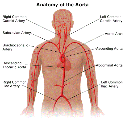
An abdominal aortic aneurysm, also identified as AAA or triple A, is a bulging, damaged location in the wall of the aorta causing in an unusual widening or ballooning greater than fifty % of the ordinary size. The aorta runs upward from the top of the left ventricle of the heart in the chest area (ascending thoracic aorta), after that curves just like a candy cane (aortic arch) downwards via the chest local area (descending thoracic aorta) into the abdomen (abdominal aorta). The aorta delivers oxygen rich blood pumped from the heart to the other parts of the body.
The most usual location of arterial aneurysm development is the abdominal aorta, mainly, the segment of the abdominal aorta directly below the renal system. An abdominal aneurysm positioned under the kidneys is named an infrarenal aneurysm.
An aneurysm may be classified
by way of its place, shape, along with reason.
The shape of an aneurysm is described as staying fusiform or saccular
which helps to recognize a true aneurysm. The more common fusiform
shaped aneurysm bulges or balloons out on all sides of the aorta. A
saccular shaped aneurysm bulges or balloons out only on one side.
A pseudoaneurysm, or untrue aneurysm, is an enhancement of just the outer part of the blood vessel wall structure. A false aneurysm may possibly be the influence of a prior surgical procedures or trauma. In some cases, a split can easily happen on the inside layer of the vessel causing in blood filling in between the layers of the blood vessel wall making a pseudoaneurysm.
The aorta is under steady tension as blood is thrown from the heart.
With every single heart beat, the walls of the aorta distend (broaden)
and then recoil (spring back again), exerting constant tension or
tension on the already weakened aneurysm wall. For that reason, there is
a potential for rupture (bursting) or dissection (split up of the
layers of the aortic wall) of the aorta, which could trigger
life-threatening hemorrhage (out of control blood loss) along with,
possibly, loss of life. The larger the aneurysm becomes, the greater the
chance of rupture.
Because an aneurysm could keep going to increase in sizing, alongside
with accelerating weakening of the artery wall, medical treatment might
be necessary.
Avoiding rupture of an aneurysm is 1 of the goals of therapy.
What triggers an abdominal aortic aneurysm to establish?
An abdominal aortic aneurysm might be triggered by multiple issues which
outcome in the breaking down of the well-organized basique components
(aminoacids) of the aortic wall that provide help as well as steady the
wall surface. The exact reason is not fully recognized.
Coronary artery disease (a build-up of plaque, which is a deposit of
fatty substances, cholesterol, cellular waste products, calcium, and
fibrin in the inner lining of an artery) is thought to perform an
important factor in aneurysmal disease, including the danger aspects
associated with vascular disease, such as:
• age (higher than 60)
• male (occurrence in males is four to five times larger compared to
that of women)
• family heritage (first level family members such as daddy or brother)
• genetic aspects
• hyperlipidemia (elevated fats in the blood)
• hypertension (high blood pressure)
• smoking
• diabetes
Some other illnesses that might cause an abdominal aneurysm contain:
• genetic disorders of connective tissue (abnormalities that can affect
tissues such as bones, cartilage, heart, and blood vessels), such as
Marfan syndrome, Ehlers-Danlos syndrome, Turner's syndrome, and
polycystic kidney disease
• congenital (present at birth) syndromes, such as bicuspid aortic valve
or coarctation of the aorta
• giant cell arteritis - a disease that causes inflammation of the
temporal arteries and other arteries in the head and neck, causing the
arteries to narrow, reducing blood flow in the affected areas; may cause
persistent headaches and vision loss
• trauma
• infectious aortitis (infections of the aorta) due to infections such
as syphilis, salmonella, or staphylococcus.
These infectious conditions
are rare.
What are the actual signals of abdominal aortic aneurysms?
Abdominal aortic aneurysms might be asymptomatic (without signs or
symptoms) or symptomatic (along with symptoms).
About 3 of every four abdominal aortic aneurysms are asymptomatic and
may be observed upon scheduled physical examination by the finding of a
pulsating mass in the abdomen. An aneurysm could also be identified by
x-ray, computed tomography scan (CT scan), or magnetic resonance imaging
(MRI) that is being done for other conditions. Because abdominal
aneurysm could be existing without signs or symptoms, it is known as
the "silent killer" - due to the fact it might possibly crack ahead of
being identified.
Pain is the most typical symptom of an abdominal aortic aneurysm. The
pain associated with an abdominal aortic aneurysm could be positioned in
the abdomen, chest, lower back, or groin area. The pain could be severe
or dull. The occurrence of pain is usually connected with the imminent
(about to happen) rupture of the aneurysm.
Acute, sudden onset of severe pain in the back and/or abdomen could
signify rupture and is a life threatening medical urgent situation.
The symptoms of an abdominal aortic aneurysm could resemble other
medical conditions or problems. Always talk to your own medical doctor
for more info.
How are aneurysms identified?
In addition to a total health-related history and physical evaluation,
analysis procedures for an aneurysm could include any, or a combination,
of the following:
• computed tomography check (Also called a CT or CAT scan) - a analysis
image procedure that utilizes a mixture of x-rays and computer system
engineering to produce cross-sectional graphics (often called pieces),
both horizontally and vertically, of the body. A CT check shows detailed
images of any part of the body, including the bones, muscles, fat, and
bodily organs. CT scans are more complete than normal x-rays.
• magnetic resonance imaging (MRI) - a analytical procedure that
utilizes a combo of large magnets, radiofrequencies, and a pc to produce
comprehensive images of body parts and systems within the body.
• ultrasound - uses high-frequency sound waves and a computer to create
pictures of blood vessels, tissues, and organs. Ultrasounds tend to be
used to view internal organs as they work, and to determine blood flow
via various vessels.
• arteriogram (angiogram) - an x-ray photo of the blood vessels used to
consider various problems, such as aneurysm, stenosis (narrowing of the
blood vessel), or blockages. A coloring (contrast) will be inserted
through a thin flexible tube placed in an artery. This dye tends to make
the blood vessels visible on x-ray.
Treatment intended for abdominal aortic aneurysms:
Unique treatment will certainly be determined by your medical doctor
based on:
• your age, overall health, and medical history
• extent of the disease
• your signs and symptoms
• your tolerance of specific medications, procedures, or therapies
• expectations for the course of the disease
• your opinion or preference treatment could contain:
• routine ultrasound procedures - to keep an eye on the size and level
of development of the aneurysm
• controlling or changing threat issues - actions such as quitting using
tobacco, managing blood sugars if diabetic, dropping bodyweight if over
weight or obese, and controlling diet fat intake may help to manage the
progression of the aneurysm
• medication - to handle variables such as hyperlipidemia (elevated
levels of fats in the blood) and/or high blood pressure
• surgery :
• abdominal aortic aneurysm open repair - a large cut is done in the abdomen to straight visualize the abdominal
aorta and repair the aneurysm. A cylinder-like tube called a graft could
be used to repair the aneurysm. Grafts are created of various
components such as Dacron (textile polyester synthetic graft) or
polytetrafluoroethylene (PTFE, non-textile synthetic graft). This graft
is sewn to the aorta, linking one end of the aorta at the site of the
aneurysm to the other end.
The open recovery is thought to be the
medical standards for an abdominal aortic aneurysm repair.
o endovascular aneurysm restoration (EVAR)
EVAR is a technique that demands just small cuts in the genitals along
with the use of x-ray guidance and specially-designed instruments to fix
the aneurysm. Through the use of special endovascular devices and x-ray
images for guidance, a stent-graft is placed via the femoral artery and
advanced up into the aorta to the site of the aneurysm.
Asymptomatic aneurysms could not demand medical intervention till they
achieve a certain dimension or are observed to be improving in size over
a specific period of time.
Ranges regarded when doing medical options
include, but are not limited to, the following:
• aneurysm size greater than 5 centimeters (about two inches)
• aneurysm growth rate 0.5 centimeters (slightly less than one-fourth
inch) over a period of six months to one year
• patient's ability to tolerate the procedure
For symptomatic aneurysms, instant intervention is recommended.
What is aortic dissection?
An aortic dissection begins with a split in the inside layer of the
aortic wall structure. The aortic wall is made up of 3 levels of tissue.
When a split occurs in the inner level of the aortic wall, blood is
then routed into the wall of the aorta, splitting the layers of tissues.
This provides extraordinary tension in the aortic wall with a potential
to rupture (burst open). Aortic dissection can be a life-threatening
urgent situation.
What causes aortic dissection?
The cause of aortic dissection is still under investigation. However,
several risk factors associated with aortic dissection include, but are
not limited to, the following:
• hypertension (high blood pressure)
• connective tissue disorders, such as Marfan's disease, Ehlers-Danlos
syndrome, and Turner's syndrome
• cystic medial disease (a degenerative disease of the aortic wall)
• aortitis (inflammation of the aorta)
• atherosclerosis
• existing thoracic aneurysm
• bicuspid aortic valve (presence of only two cusps, or leaflets, in the
aortic valve, rather than the normal three cusps)
• trauma
• coarctation of the aorta (narrowing of the aorta)
• hypervolemia (excess fluid or volume in the circulation)
• polycystic kidney disease (a genetic disorder characterized by the growth of numerous cysts filled with fluid in the kidneys)
What are the symptoms of aortic dissection?
The most commonly reported symptom of an acute aortic dissection is
severe, constant pain, sometimes described as "ripping" or "tearing"
and located in the chest, the middle of the abdomen, the lower back, or
the pelvis area. The pain may be "migratory" moving from one place to
another, according to the direction and extent of the dissection.
The symptoms of aortic dissection may resemble other medical conditions
or problems. Always consult your physician for more information.
How is aortic dissection diagnosed?
In addition to a complete medical history and physical examination,
diagnostic procedures for an aortic dissection may include any, or a
combination, of the following:
• computed tomography scan (Also called a CT or CAT scan.) - a
diagnostic imaging procedure that uses a combination of x-rays and
computer technology to produce cross-sectional images (often called
slices), both horizontally and vertically, of the body. A CT scan shows
detailed images of any part of the body, including the bones, muscles,
fat, and organs. CT scans are more detailed than general x-rays.
• transesophageal echocardiogram (TEE) - a diagnostic procedure that
uses echocardiography to assess the heart's function and structures. A
transesophageal echocardiogram is performed by inserting a probe with a
transducer down the esophagus. By inserting the transducer in the
esophagus, TEE provides a clearer image of the heart because the sound
waves do not have to pass through skin, muscle, or bone tissue.
The physician will determine the most appropriate examination. When a
diagnosis of aortic dissection is confirmed, immediate intervention is
necessary. Medical intervention or surgery will be required depending on
the location of the aortic dissection.
Learn more about abdominal aneurysm and aortic dissection.
